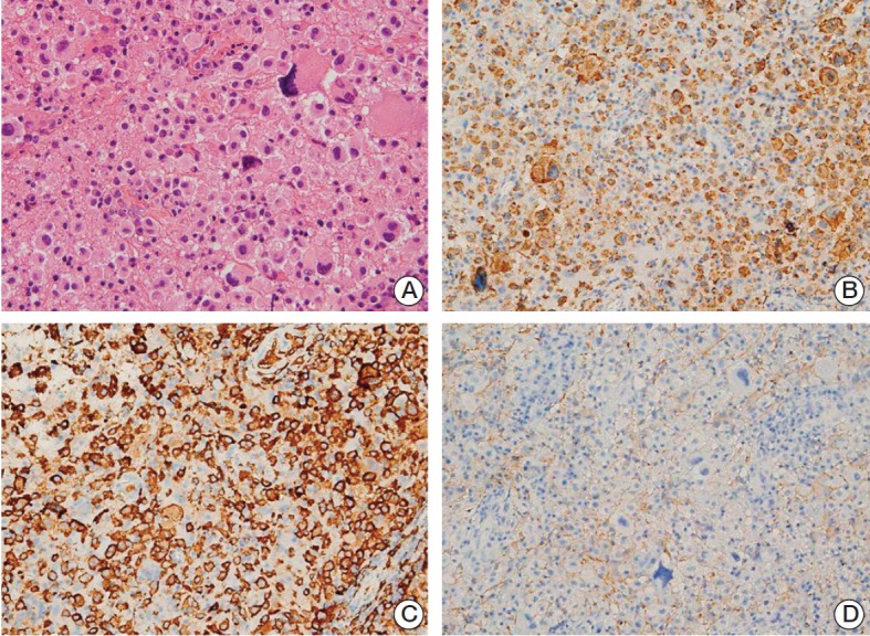
Fig. 3.

Histologic features of left parieto-occipital lesion from the second biopsy. (A) Large cells were arranged in a solid sheet, with a few intermixed larger bizarre cells, similar to findings in the first biopsy specimen. The nuclei of the background cells showed slight pleomorphism and coarse chromatin with no or small nucleoli. The cytoplasm was eosinophilic and cell membranes were not well-defined (H&E staining, ×400). (B-D) The cells were positive for CD68 (B) and CD163 (C), but negative for glial fibrillary acidic protein (D), consistent with histiocytic differentiation (×200).
