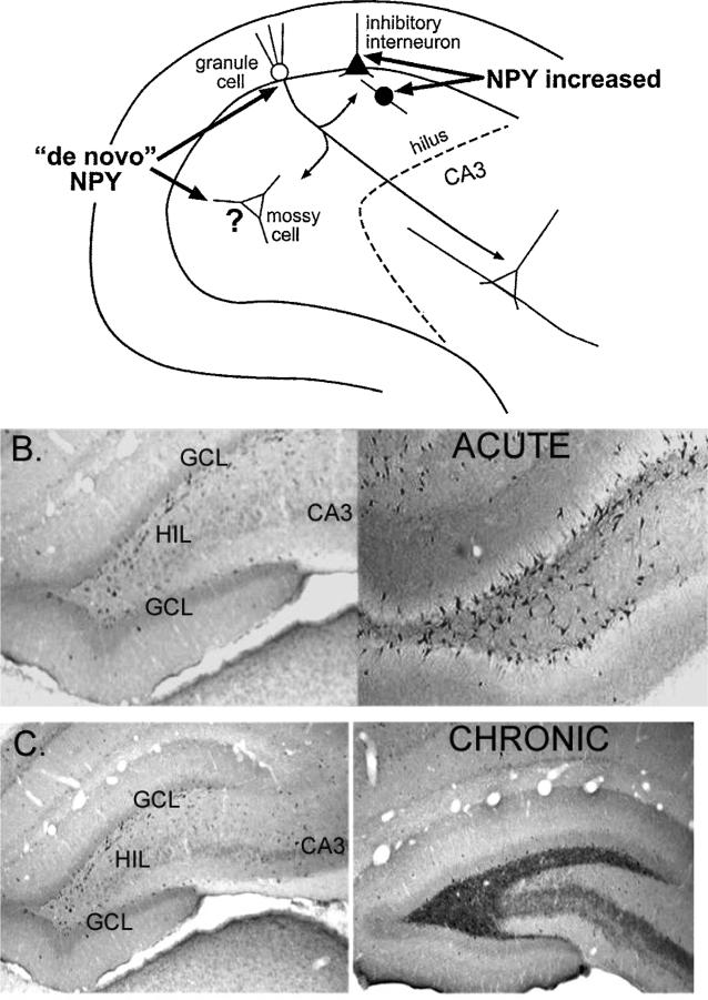
Figure 5.
Changes in NPY expression after seizures. A. A diagram is used to illustrate where NPY protein increases in the adult rat dentate gyrus after seizures. It appears to increase in inhibitory neurons, and also develop in neurons such as granule cells and mossy cells that do not normally express the protein (de novo expression). B. The increase in NPY protein in the dentate gyrus is shown using an antibody to NPY. Left: Saline control. Right: 1 day after status epilepticus. C. Increased NPY in mossy fibers is illustrated after chronic seizures. Left: Saline control. Right: 2 months after status epilepticus. Spontaneous seizures were observed in the animal that had status epilepticus.

