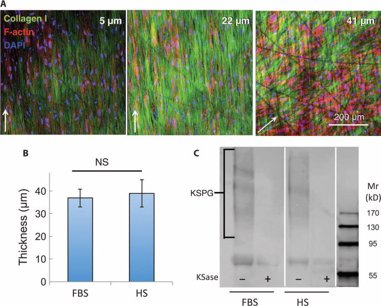
Fig. 3. Generation of a stroma-like three-dimensional matrix ex vivo.

ECM produced by LBSCs cultured on aligned nanofiber substrate for 4 weeks was imaged by confocal microscopy capturing optical sections at different z levels above the substratum. (A) Type I collagen fibrils (green) and keratocytes (nuclei, blue; F-actin, red) are shown at different depths of the construct. (B) Thickness of collagenous matrix at 4 weeks in HS or FBS was determined from confocal analysis. Data in (B) show averages ± SD from cell lines from four different donors. Lack of significance (NS; P > 0.05) was determined by a two-sided t test. (C) Cornea-specific keratan sulfate proteoglycan (KSPG) was detected by immunoblotting. Alternate lanes show sensitivity of the heterogeneous KSPG band (130 to 300 kD) to keratanase (KSase). Mr, relative molecular mass.
