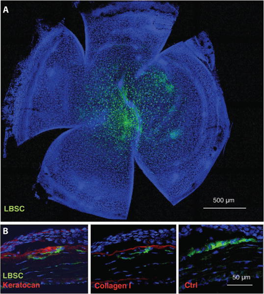
Fig. 4. LBSC engraftment and stromal matrix synthesis in mouse cornea in vivo.

Fluorescent DiO-labeled human LBSCs were transferred to a superficially debrided mouse cornea in a fibrin gel as depicted in fig. S3. (A) One week after wounding, whole-mount staining showed persistence of the human LBSCs (green) in the central corneal region. (B) At 1 month, histological sections immunostained with human-specific antibodies show human keratocan and collagen type I. Omission of primary antibody controlled nonspecific staining. In all images, nuclei are stained with 4′,6-diamidino-2-phenylindole (DAPI) (blue). Anterior of the eye is oriented up in each image in (B), and the corneal epithelium is visible as a dense layer of cells near the top of each image. Ctrl, control.
