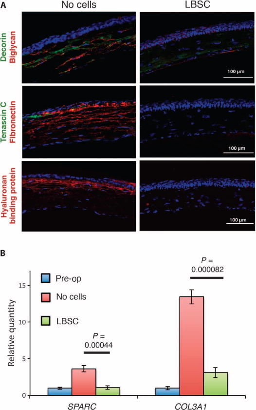
Fig. 5. LBSCs block deposition of fibrotic matrix in healing murine corneas.

(A) Debridement-wounded mouse corneas were treated with fibrin gel only (no cells) or with 50,000 LBSCs in fibrin gel. After 4 weeks of healing, histological sections (epithelium oriented up) were stained for fibrotic markers decorin, biglycan, tenascin C, fibronectin, and hyaluronan. Images are representative of sections from three corneas for each condition. (B) Quantification of SPARC and type III collagen (COL3A1) mRNA pre-operative (Pre-op) and 2 weeks after treatment with LBSCs or no cells. Data are averages ± SD (n = 3). P values were determined by a two-sided t test.
