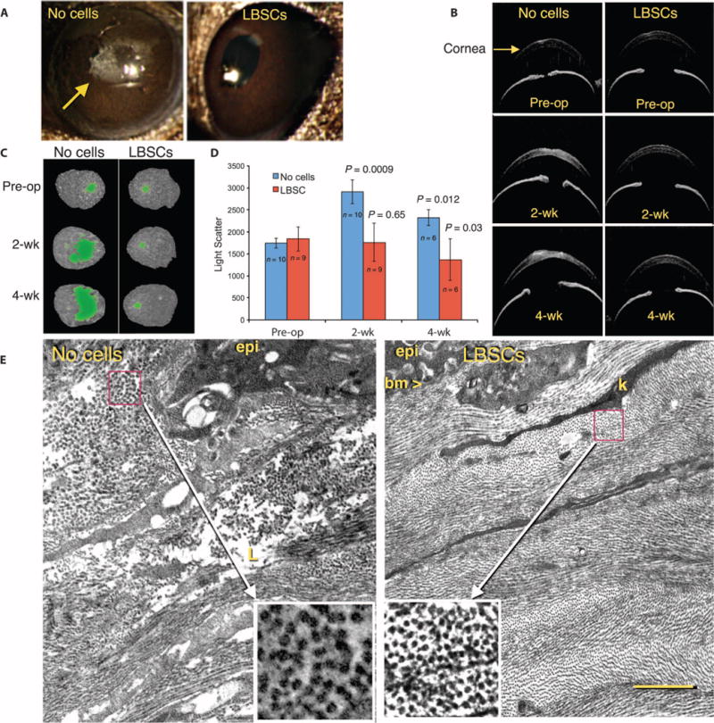
Fig. 6. LBSC treatment influences light transmission properties of ECM deposited after debridement.

(A) Macroscopic images of mouse eyes in diffuse lighting reveal opaque scars (arrow) in untreated (no cells) eyes but none in LBSC-treated corneas. (B) OCT imaging shows transverse optical sections of preoperative (Pre-op) eyes and those 2 and 4 weeks after debridement, with scarring visible as bright pixels in the corneal stroma. (C) Thresholding of high-intensity pixels in three-dimensional (3D) OCT images of individual corneas defines scarred region (green) at 2 and 4 weeks after debridement. (D) Light scatter in 3D OCT scans at 2 and 4 weeks was compared with the values in preoperative eyes. Data are means ± SD. The number of eyes is indicated in the graph. P values were determined with unpaired t tests at each time point compared to respective Pre-op values (table S4). (E) Transmission electron micrographs 4 weeks after debridement show the ablated region of the anterior stroma. epi, epithelial cells; bm, basement membrane; k, keratocyte processes; L, amorphous matrix deposit (lake). Insets show magnification of a box (1 μm × 1 μm) from the indicated region containing orthogonal views of collagen fibrils. Scale bar, 2 μm. Images are representative of n = 3 animals.
