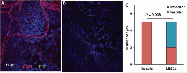
Fig. 7. Vascularization of debridement wounds.

One month after wounding, whole-mount corneas were stained with antibody to CD31 (red) to detect ingrowth of blood vessels. Cell nuclei were imaged with DAPI (blue), and added human LBSCs appear green. (A) Vessels in a healed cornea in which no cells were added. (B) DiO-labeled LBSCs are visible (green), but no vessels were present in the central LBSC-treated wound. (C) A stacked bar graph shows the proportion of vascularized corneas in human LBSC-treated (n = 5) and untreated (n = 5) mouse eyes analyzed by staining as in (A). P value was obtained from a two-tailed χ2 test with a 2 × 2 contingency table.
