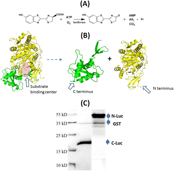
Fig 4. Split fragments of luciferase.
(A) Luciferase assay. (B) Split luciferase to give two complementary fragments N-Luc 2–416 and C-Luc 398–550, each his-tagged at N terminus and C terminus respectively (the structure is from PDB ID 2D1S) [39]. (C) SDS-PAGE picture of N-Luc and C-Luc.

