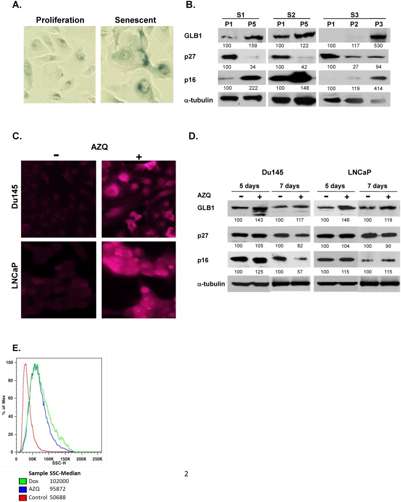
Fig 2. GLB1 is increased in replicative and therapy-induced senescent (TIS) cell culture models.
(A) Human prostate epithelial cells were cultured to senescence by repeated cell passaging demonstrate an enlarged, more complex senescent cell morphology and increased β–gal staining in passage 5 (P5) than in proliferating (P1) cultures. (B) Western blot comparing proliferating HPECs (P1) versus later passages (P5) for separate cultures (S1, S2, S3) are shown. GLB1 and p16 expression is increased in senescence. Cultures contained >50% senescent cells by SA-βgal staining. Images were normalized to α-tubulin. (C) GLB1 mmunofluorescence increases in cancer cells displaying TIS. Du145 and LNCaP cells were treated with 250nM AZQ or DMSO (control) for 3 days, kept in drug-free media for an additional two days, then stained with mouse anti-human GLB1 antibody and detected with anti-mouse-Alexa 647. (D) Western blot analysis of GLB1 expression at 5 or 7 days after AZQ treatment performed in DU145 and LNCaP PCa lines. Cells were compared against a DMSO-treated control group (Con) and normalized to α-tubulin expression levels. (E) Cell granularity and complexity analysis of senescence by flow cytometry. Du145 cells were treated with AZQ or Doxorubicin for 3 days, kept in drug-free media for two days then collected and applied to MACSQuant flow cytometer immediately. Side scatter (SSC), which increases in senescent cells, increased in senescent AZQ and Doxorubicin treated cells compared to control.

