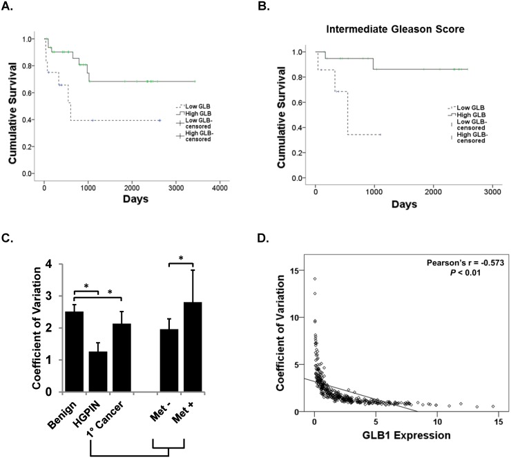
Fig 4. Increased GLB1 identifies patients at decreased risk of PCa recurrence.
GLB1 expression on a per-core basis was measured using the Vectra platform in all tissues. Patients were stratified into high or low GLB1 levels based on median core expression. Kaplan-Meier survival curves are shown. (A) Increased GLB1 expression is associated with longer PSA-free recurrence in PCa patients. Stratified by low (n = 12) and high GLB1(n = 32) expression, cumulative PSA-free survival significantly increases in patients who had high GLB1 levels in a log rank analysis (p = 0.025). (B) High (n = 20) and low (n = 7) GLB1 levels predict PSA-free survival times (629 vs. 2314 days, respectively; p = 0.013) in intermediate Gleason Score (GS 6–7) patients. (C). Per cell GLB1 expression is more homogenous in lower grade PCa tissues which express increased GLB1. GLB1 expression on a per-cell basis was then measured using the Vectra platform in all tissues. HGPIN had the least heterogeneity (p<0.001). Primary cancer tissue overall has significantly less heterogeneity than benign (p<0.001). Primary cancer cells with associated metastatic disease (Met+) had lower levels compared to localized tumors without the presence of metastatic (Met-) disease (p = 0.001) (D). Mean nuclear GLB1 expression was inversely correlated with the coefficient of variation (representing heterogeneity) of the cores (Pearson's r = -0.573; p<0.01; N = 265). Tissues that display the highest levels of GLB1 staining have the most homogenous staining.

