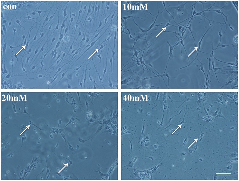
Fig 1. The morphology of SGNs in different interventions in stereoscopic microscope.
Glu and two inhibitors were added to the SGNs in integrated medium. In addition to the control group, the drugs that were added to the cultured SGNs included 20 mM Glu, 20+PD150606 and 20+Z-VAD-FMK. Morphological changes were clearly observed in the four groups. The neurites of the SGNs were counted by Image-Pro Plus IPP with the scales. Scale bar = 100 μm.

