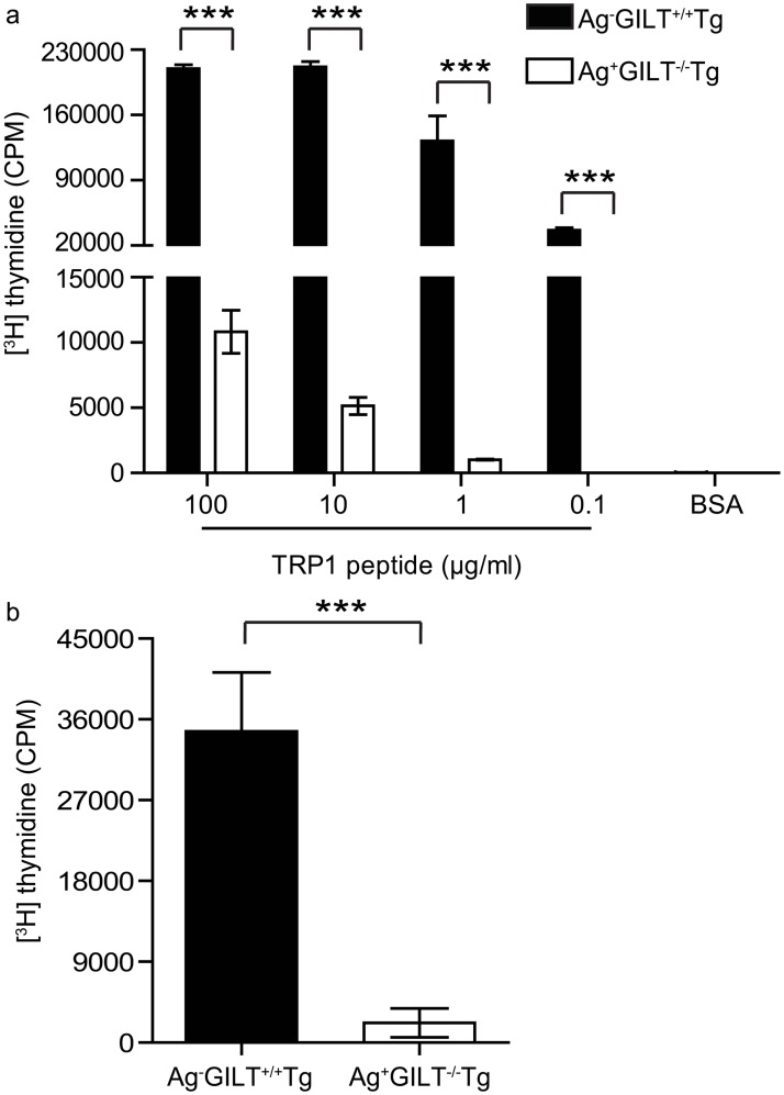
Fig 2. Melanoma-specific T cells from TRP1-expressing mice undergo diminished proliferation in response to TCR stimulation.
CD4+ lymph node cells from Ag-GILT+/+Tg and Ag+GILT-/-Tg mice were cocultured with irradiated T cell-depleted splenocytes from wild-type mice and (a) TRP1 peptide or (b) anti-CD3 and anti-CD28 antibodies for five days. Columns and bars represent means ± standard error of triplicate samples from one experiment. Data are representative of two independent experiments and compared using an unpaired t test (***p<0.001).

