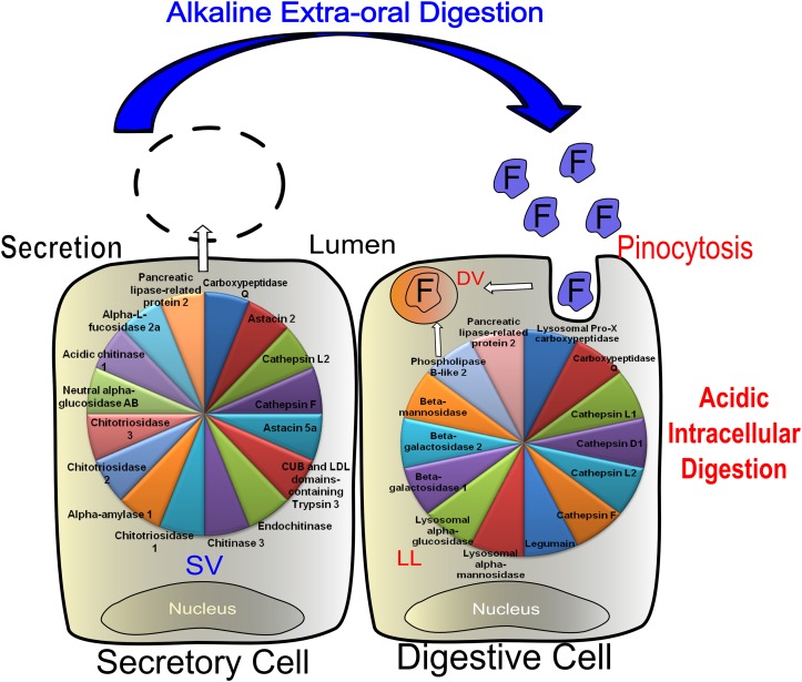
Fig 8. Schematic representation of midgut and midgut glands secretory (SC) and digestive cells (DC).
Figure displays enzymes present in secretory vesicles (SV) and lysosome-like (LL) organelles. Lysosomes probably fuse or exchange contents with pinocytic vesicles to end up in digestive vacuoles. DC: digestive cells, DV: digestive vacuoles, F: pre digested food, M: mitochondria, P: pinocytosis, RER: rough endoplasmic reticulum, S: spherites, SC: secretory cells.

