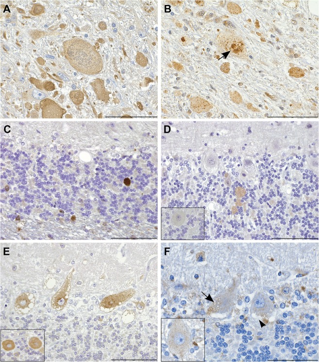
Fig 4. Immunohistochemistry indicates disturbed autophagic flow in neurons.
(A) Axonal spheroids stain diffusely positive for LC3B. IHC LC3B, scale bar 100 μm. (B,C) The granular cores of the spheroids are positive for (B) ubiquitin (arrow) and (C) p62. IHC ubiquitin and p62, scale bars 20 and 100 μm, respectively. (D) Smooth axonal swellings in the cerebellar cortex contain ATG4D. Inset: control. IHC ATG4D, scale bar 100 μm. (E) Affected neurons show increased perinuclear granular LC3B positivity. Inset: control. IHC LC3B, scale bar 100 μm. (F) Neuronal vacuoles are partially LAMP2 positive (arrow) and partially negative (arrow head). Inset: control. IHC LAMP2, scale bar 20 μm.

