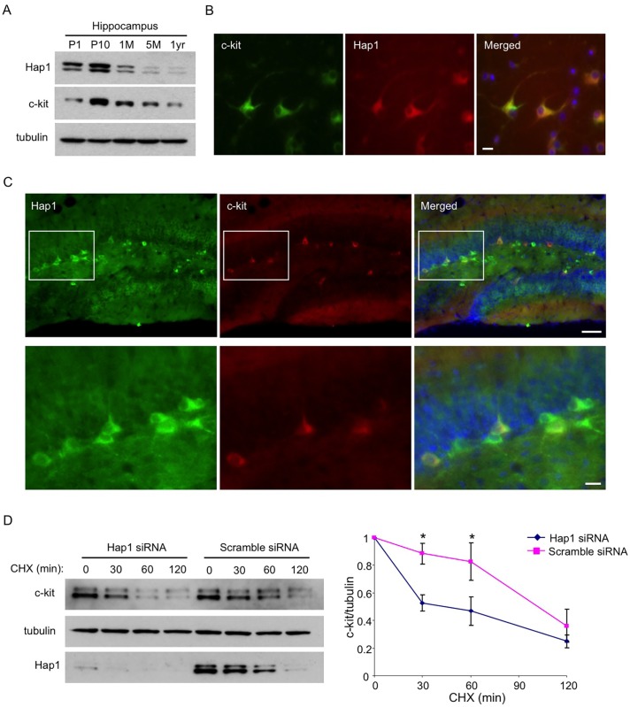
Fig 6. Hap1 is coexpressed with c-kit and stabilizes its level in vitro.
(A) Age-dependent expression levels of Hap1 and c-kit in the hippocampus of mice at different ages. Both proteins peak in their expression at the postnatal stage. (B) Hippocampal neuronal culture at DIV5 was immunostained with antibodies against Hap1 and c-kit. The two proteins are coexpressed in the majority of cultured hippocampal neurons. Scale bar: 10 μm. (C) Brain sections from P15 WT mouse were immunostained with antibodies to Hap1 and c-kit. Hap1 and c-kit are coexpressed in some neurons in the hippocampal DG. Lower panel represents magnified images of the boxed areas in the upper panel. Scale bars: 40 μm (upper panel), 10 μm (lower panel). (D) Plasmid construct expressing c-kit was transfected into Neuro2A cells that had been treated with either scramble or Hap1 siRNA. Protein stability of c-kit was then assessed by cycloheximide (CHX) chase followed by western blot (left panel). Quantification of 3 independent experiments is shown on the right panel. Hap1 knockdown markedly decreased the half-life of c-kit in Neuro2A cells. Error bars represent SEM. *p<0.05.

