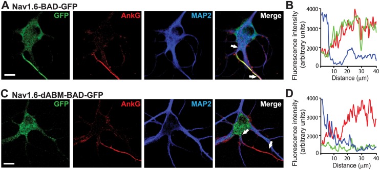
Fig 2. Nav1.6-BAD-GFP but not Nav1.6-dABM-BAD-GFP co-localizes with axonal markers.
(A,C) Compression of confocal z-stacks of DIV8 rat hippocampal neurons co-transfected with ankG-mCherry and either wild-type Nav1.6-BAD-GFP or Nav1.6-dABM-BAD-GFP and immunolabled for MAP2. A) Nav1.6 is enriched within the ankG positive AIS. B) Line profile of the AIS showing the simultaneous increase of Nav1.6 (green) and ankG (red) and the decrease of MAP2 (blue) with increasing distance from the soma. C) Nav1.6-dABM-BAD-GPF is found throughout the somatodendritic region of the neuron, but is not enriched in the ankG positive AIS. D) Line profile of the AIS showing the increase in ankG (red) while the expression levels of the mutant Nav1.6 (green) and MAP2 (blue) are low within the AIS. Scale bars represent 10 μm.

