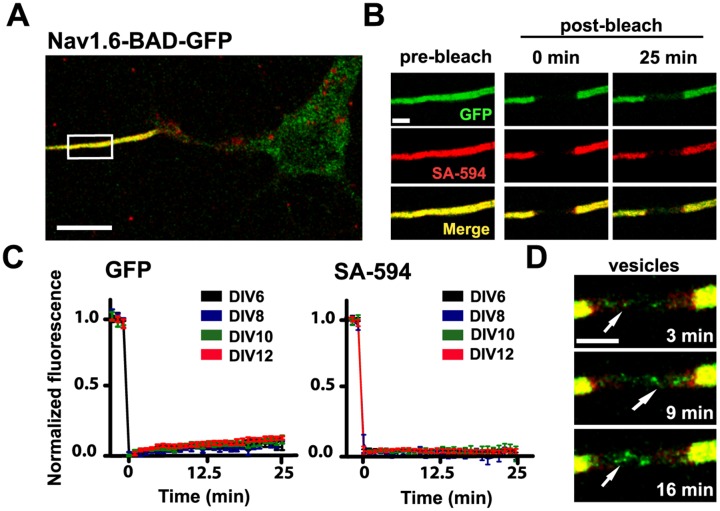
Fig 4. Nav1.6 is stable in the AIS of mature neurons.
A) A representative compressed confocal z-stack of a DIV10 rHN expressing Nav1.6-BAD-GFP. The high density of Nav1.6 in the AIS is labeled both by the GFP fluorescence (green) and by surface labeling of the BAD tag via SA-594 (red). B) Enlargement of the white box in (A) showing fluorescence before photobleaching, immediately after photobleaching and 25 min postbleach. C) Average normalized FRAP curves over 25 min for the GFP fluorescence and surface-specific SA-594 fluorescence. Days post-transfection are indicated. On average, the GFP recovered 7.5±0.2%, n = 8, for DIV6 and 9.7±1.8%, n = 6, for DIV10 mean ± s.e.m.)Images were acquired every minute to minimize photobleaching during the recovery. D) Detection of mobile GFP-containing trafficking vesicles within the bleached AIS. The different time points illustrate the detection of dynamic puncta (arrows) at the indicated postbleach times. Scale bars represent 10 μm (A) or 2 μm (B,D).

