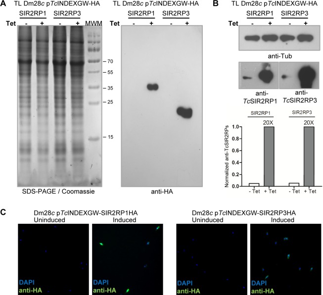
Fig 2. Inducible expression of sirtuins in epimastigotes.
Equal amounts of parasite total lysate from each line (pTcINDEXGW-SIR2RP1HA and pTcINDEXGW-SIR2RP3HA) in the absence (-) or presence (+) of 0.25 μg/ml Tetracycline for 24 hours, were loaded on SDS-PAGE and stained with Coomassie (left panel), followed by western blot analysis using rat anti-HA monoclonal antibodies (A), or mouse anti-Tubulin and specific rabbit polyclonal antibodies against TcSIR2RP1 and TcSIR2RP3 (B). The degree of overexpression observed with the specific antibodies were quantified and normalized to α-tubulin intensity. (C) Immunofluorescence microscopy of uninduced and induced (0.25 μg/ml Tetracycline, 24 hours) parasites using rat anti-HA and FITC-conjugated anti-rat antibodies (green). DNA was stained with DAPI (blue).

