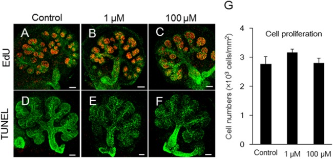
Fig 3. Effects of melatonin on both proliferation and apoptosis during submandibular gland branching morphogenesis.

Growing SMG cells in control, 1 μM melatonin, and 100 μM melatonin-treated groups were positive for EdU (A, B, C) and for TUNEL (D, E, F) as markers of cell proliferation and apoptosis, respectively. Proliferating cell nuclei appeared as red punctate spots (A, B, C). Lectin peanut agglutinin (PNA)—Alexa Fluor 647 was used to stain the epithelium green (A-F). Apoptosis of epithelial cells was not detected by TUNEL in red (D, E, F). Scale bar: 100 μm. Fluorescent EdU staining in the epithelium were quantified using ImageJ software (n = 5) (G). The total EdU positive cells were expressed as a ratio of the area. Bars represent the mean ± SEM.
