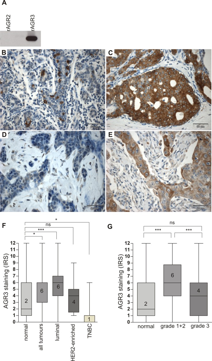
Fig 2. Up-regulation of AGR3 protein in G1/G2 grade and luminal breast cancer.
(A) Western blot detection of human recombinant AGR3 (rAGR3, 100ng) but not recombinant AGR2 (rAGR2, 100ng) by monoclonal AGR3 antibody used for IHC analysis. (B) Cytoplasmic staining of AGR3 in isolated cells of the normal breast epithelium. (C) Strong cytoplasmic AGR3 protein expression in epithelial cancer cells of an IHC-defined luminal breast tumour. (D) Absent and (E) weak cytoplasmic AGR3 expression in two different triple negative breast tumours. (F) Box plot analysis demonstrating a significant up-regulation of AGR3 in all tumours (n = 190) and the luminal subtype (n = 113), but a significant reduction of expression in the triple negative breast cancer cases (TNBC, n = 23) compared to normal breast tissues (n = 39). (G) Box plot analysis showing a highly significant up-regulation of AGR3 in G1 and G2 breast tumours (n = 104) compared to normal controls (n = 39). Horizontal lines: grouped medians. Boxes: 25–75% quartiles. Vertical lines: range, minimum and maximum. Ns: not significant, * P < 0.05, *** P < 0.001. IRS: immunoreactive score.

