
Figure 6.
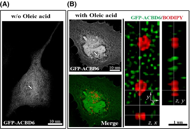
hACBD6 is not associated to LDs in HeLa cell. HeLa cells were grown on coverslips and transfected with GFP-ACBD6 (green) for 48 h (A). LDs production (B) was induced by addition of oleic acid in the growth medium for 24 h and LDs were labeled with BODIPY C12 (red). After cell fixation in 4% paraformaldehyde, DNA was stained with the Hoechst dye (blue).) Cropped orthogonal (z, x) and (z, y) views of the deconvolved Z-stack merged image is shown on the right. Note the lack of overlay of the GFP and BODIPY signal (tMCC of 0.0431). LDs, lipid droplets.