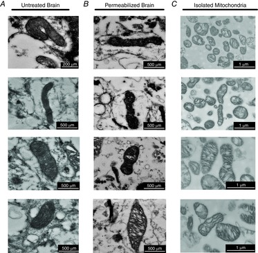
Figure 3. Imaging of mitochondrial morphology.

Sections of intact mitochondrial reticulum are present in untreated cortex (A) and following retrieval from a full-length permeabilized respiration protocol (B). C, isolated mitochondria are numerous, swollen and absent of mitochondrial networks. Scale bars represent 200–500 nm for in vivo (left) and in situ mitochondria (centre), 1 μm for isolated mitochondria (right).
