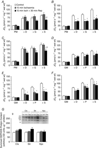
Figure 4. Application in acute ischaemia–reperfusion injuries.

Whole brain ischaemia was induced through a bilateral carotid artery occlusion for 15 min and reperfusion was permitted for 30 min (n = 6/group). Pyruvate-stimulated respiration observed in the cortex (A), striatum (B) and hippocampus (C) in control (white bars), ischaemia (grey bars) and reperfusion (black bars). Glutamate-stimulated respiration demonstrates unique dysfunction in the cortex (D), striatum (E) and hippocampus (F). G, samples retrieved from the respirometer demonstrated that protein content of select electron transport chain subunits from complexes I to V were not affected by ischaemia–reperfusion injuries. CI-V, complex I–V; n = 6 for all groups. *P < 0.05 vs. control; †P < 0.05 vs. ischaemia. ctx, cortex; D, ADP; G, glutamate; hpc, hippocampus; isch, ischaemia; M, malate; P, pyruvate; rep, reperfusion; S, succinate; str, striatum.
