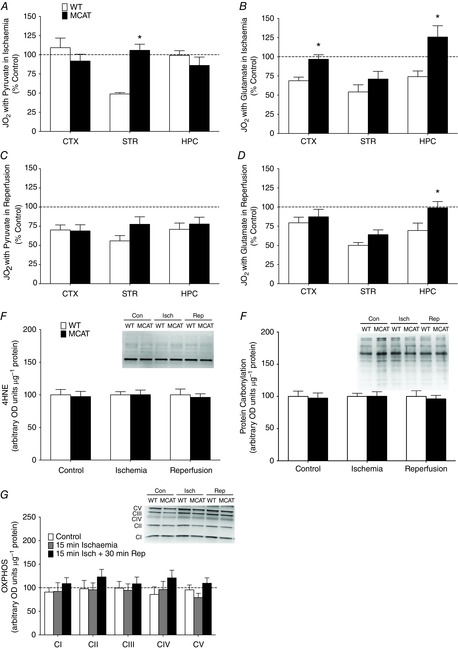
Figure 6. Simplified application when approaching mechanisms with transgenic models.

Ischaemia–reperfusion injuries were performed in mice expressing MCAT to buffer reactive oxygen species (n = 5/group). Comparing the maximal pyruvate-stimulated (A) and glutamate-stimulated (B) respiration of ischaemic cortex, striatum and hippocampus in WT and MCAT mice. Maximal pyruvate-stimulated (D) and glutamate-stimulated (E) respiration of cortex, striatum and hippocampus following ischaemia and reperfusion. Respiration values are shown as a percentage of control values within a genotype. Acute changes in function are undetectable using protein markers of oxidative damage such as lipid peroxidation (4HNE) (E) and protein carbonylation (F). G, protein content of subunits of the electron transport chain did not change in examined cortex of WT and MCAT mice in any condition. n = 5 for all groups. *P < 0.05 MCAT vs. WT percentage control under the same condition. 4HNE, 4-hydroxy-2-nonenal; con, control; ctx, cortex; hpc, hippocampus; isch, ischaemia; MCAT, mitochondrial-targeted catalyse; rep, reperfusion; str, striatum; WT, wild-type.
