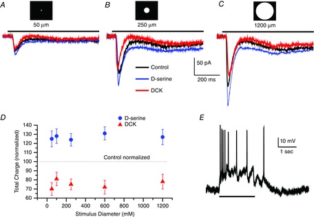
Figure 3. Increasing the stimulus spot diameter does not change the sensitivity to DCK or d-serine.

Whole-cell voltage-clamp recording (Vhold = −58 mV) of a sustained ON retinal ganglion cell in an eyecup preparation responding to a 50 μm (A), 250 μm (B) and 1200 μm (C) light stimulus 2 s in duration in control Ringer solution (black trace), d-serine (100 μm, blue trace) and DCK (30 μm, red trace). Scale bar under the trace in B applies to traces in A, B and C. D, summary data from eight cells plotting the mean ± SEM charge for responses studied for the range of diameters indicated. Recording in control Ringer solution was standardized to 100% as indicated by the dashed line. d-serine responses (blue dots) and responses recorded in DCK (red triangles). Light stimuli of 50, 100, 250, 600 and 1200 μm diameter were used to generate this data. ON and OFF responses were combined before calculating the mean. E, a current clamp record from an ON ganglion cell, light stimulus is represented by the black line at bottom.
