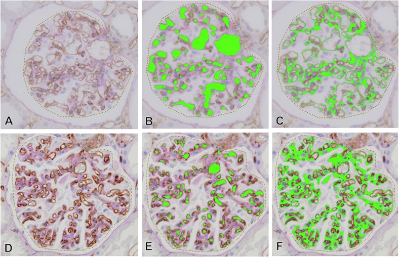
Fig 1. The areas of glomerular capillaries and glomerular ECM in computer-assessed morphometric analysis.
The area of glomerular tuft (dotted line in A and D), the area and number of glomerular capillaries (green areas in B and E), and the area of glomerular ECM (green areas in C and F) in each glomerulus were assessed by computer-assisted image analyzer. In the nearly normal glomeruli in light microscopic findings (A-C), large glomerular capillary area was noted with minimal ECM accumulation. In contrast, the area of glomerular capillaries decreased with narrowing glomerular capillaries and the accumulation of glomerular ECM in glomerulus in the development of glomerular sclerosis (D-F).

