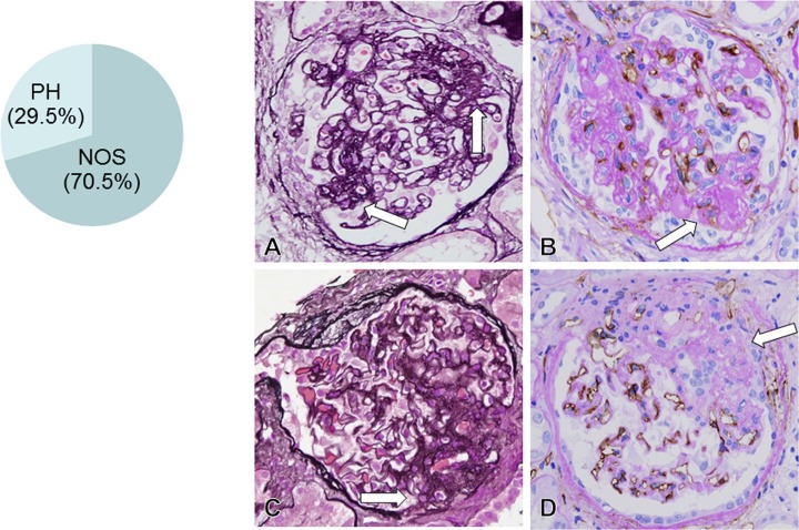
Fig 2. FSGS lesion in idiopathic membranous nephropathy(MN) (A, C: PAM stain, x600; B, D: CD34 stain, x600).
The biopsy samples from 26 MN-FSGS(+) cases included 534 glomeruli except for 55 global sclerotic glomeruli. In 534 glomeruli, 44 glomeruli (8.2%) had FSGS lesion, consisted of not otherwise specified (NOS) lesion type (31 glomeruli, 70.5%) and perihilar (PH) lesion type (13 glomeruli, 29.5%). In glomeruli with NOS type of FSGS lesion (arrow in A), FSGS lesion was noted in areas other than perihilar region, with hyalinosis and adherence lesion between tuft and Bowman’s capsule. In FSGS lesion (arrow in B), loss of CD34+ capillaries was evident with mesangial ECM accumulation. In glomeruli with PH type of FSGS lesion (arrow in C), perihilar sclerosis with hyalinosis was noted with loss of CD34+ glomerular capillaries (arrow in D) and mesangial ECM accumulation.

