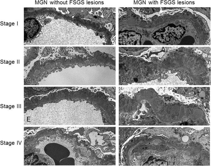
Fig 8. The alteration of glomerular capillaries in cases of idiopathic membranous nephropathy (MN) with/without FSGS lesion.
Representative alterations of glomerular capillary walls were indicated from stage I to Stage IV of MN. The thickening of glomerular capillary walls was seen with ECM accumulation in both MN cases with and without FSGS lesion, with subepithelial deposits in stage I, spike formation in stage II to III, and wash out of deposits in stage IV. However, in MN-FSGS(−) cases, glomerular capillary walls with subepithelial deposits in stage I to IV were characterized by well-preserved fenestra of glomerular endothelial cells, less widening of subendothelial space, mild thickening of glomerular capillary walls and GBM, and relatively preserved foot processes of podocytes. On the other hand, in MN-FSGS(+) cases there were prominent thickening of glomerular capillary walls with ECM accumulation, loss of foot processes of podocytes, and endothelial cell injury, indicated by loss of fenestra, swelling of cytoplasm and dilatation of subendothelial space.

