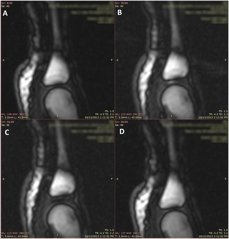
Fig 3. Still frames from a representative trial of joint cracking in the same MCP joint.
The right 4th MCP joint in the resting phase (A). The MCP joint as seen during distraction of the MCP joint in the frame just prior to joint cracking / joint separation (B). The MCP joint visualized in the next frame immediately after joint cracking (C). The joint in the refractory phase immediately after removal of distraction forces (D).

