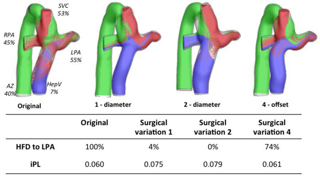
Figure 8.
Surgical variations for Patient H that resulted in the highest differences from the original configuration (left); Surgical variation 1 represents a decrease in the FP baffle (−2mm), and Surgical variation 2 an increase (+2mm); Surgical variation 4 is a change in offset. Table shows the HFD and iPL values. Streamtraces are color coded by vessel of origin: hepatic vein (blue), SVC (red), AZ (green).

