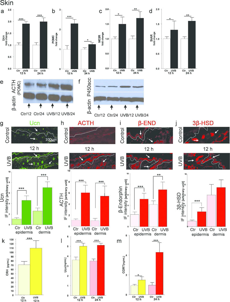
Figure 3.
Cutaneous equivalent of the HPA axis in C57BL6 mice is stimulated upon UVB radiation.
Expression of genes coding Ucn (a), POMC (b), MC2R (c) and StAR (d) after UVB exposure compared to control (shame-treated) animals. Data presented as fold change ± SD. Protein estimation with Western Blot for ACTH/POMC (e) and P450scc (f). In situ expression of Ucn (g), ACTH (h), β-END (i) and 3β-HSD (j) antigens measured by immunofluorescence with corresponding quantification of immunopositive signal intensity (inserts to the subpannels). Arrows indicate examples of positive signals. ELISA evaluation of peptide CRH (k), Ucn (l) and steroid CORT (m) concentrations. Data are presented in pg or ng/mL per 4 µg of total proteins extracted, and analyzed using Student’s t-test, * p<0.05, ** p<0.01, and *** p<0.001.

