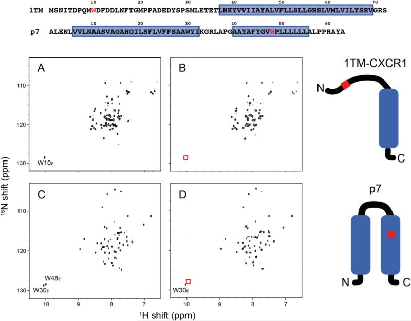
Figure 4.
Comparison of 1H-15N HSQC spectra of uniformly 15N-labeled membrane protein constructs in DHPC micelles. (A) Wild-type 1TM-CXCR1. (B) W10HQA 1TM-CXCR1. (C) Wild-type p7. (D) W48HQA p7. Absence of Trp indole NHε signals shown in red boxes indicates complete incorporation of unnatural amino acid HQA into residue Trp 10 in 1TM-CXCR1 and into residue Trp 48 in p7. The mutation site is colored in red in the amino acid sequences and schematic drawings of proteins. Note that the amide chemical shifts for wild-type and HQA-incorporated proteins are identical except in the vicinity of the mutated site.

