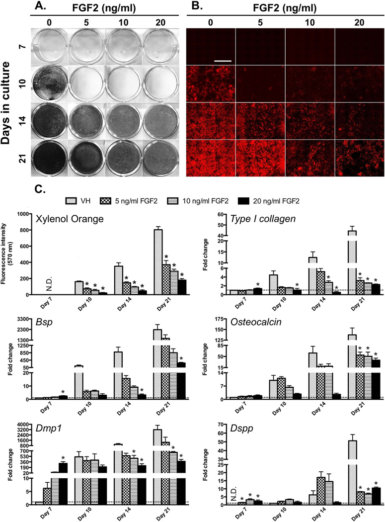
Figure 1. Concentration-dependent effects of FGF2 on mineralization and the expression of markers of mineralization and dentinogenesis in primary dental pulp cultures.
(A) Representative images of von Kossa-stained dishes.
(B) Representative composite of 5× scanned images of XO-stained live cultures at various time points. The magnifications of all micrographs are identical. Scale bar = 2 mm.
(C) Histogram showing the changes in the intensity of XO fluorescence and in the levels of expression of various markers. The intensity of XO staining is expressed as absolute values and the expression levels of markers of mineralization and dentinogenesis are expressed as relative values. Expression of all mRNAs except Dspp is normalized to VH at day 7, which is arbitrarily set to 1 and indicated by the dashed line. The expression of Dspp is normalized to VH at day 10, which is arbitrarily set to 1 and indicated by the dashed line. Results in all histograms represent mean ± SEM of at least three independent experiments; *p ≤ 0.05 relative to VH at each time point. N.D. = not detected. Note the increases in the expression of markers of mineralization and dentinogenesis at day 7 followed by decreases in their expression at days 10–21 in FGF2-treated cultures as compared to control.

