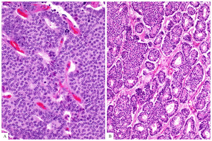
Figure 1.
Well differentiated neuroendocrine tumors may reveal different growth patterns (a-diffuse and b-glandular growth patterns are depicted here). The cells vary in size but usually have moderate amount of eosinophilic cytoplasm and nuclei are uniform in size and shape. Mitotic figures are rare (by definition, between 2 and 20 per 10 HPF for Grade 2).

