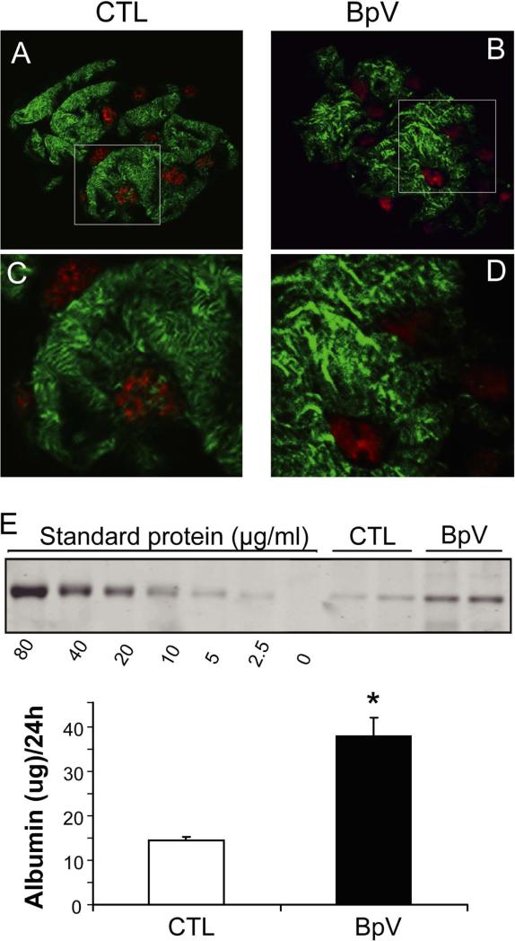
Figure 3. In mice, PTEN inhibition causes podocyte cytoskeletal rearrangement and albuminuria.
A and B: After C57B6 mice were injected with saline (CTL, A) or BpV (ip, 1mg/Kg, C) for 1h, preparation of glomeruli, and confocal immunoflorescence microscopy, were performed as described in Methods. F-fibers in podocytes were visualized with phalloidin (Green) and podocyte nuclei were labeled with WT1 (red). C and D: enlarged images from the areas indicated in the upper panels.
E: C57B6 mice were injected with BpV (ip, 1mg/Kg/12 h, two dosages) and 24h urines were collected. Urine samples were subjected to SDS electrophoresis and the amount of albumin determined as described in Methods.

