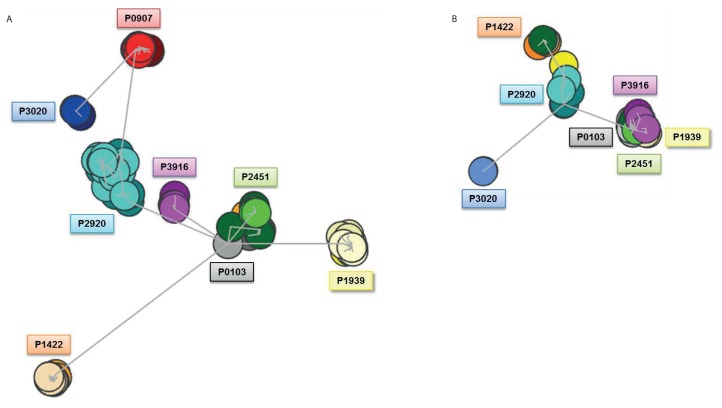
Figure 3.
Composite clusters of subset #4 IG sequences at the nucleotide level. Figure 3A illustrates cluster formation following analysis of the IGHV–IGHD–IGHJ nucleotide sequences (n = 511). Seven distinct clusters were observed; six clusters represented a single patient each, while P0103 and P2451 remained clustered together, thus accounting for the seventh cluster. Figure 3B highlights cluster formation following analysis of the IGKV–IGKJ nucleotide sequences (n = 397) and highlights the distancing of the two central cores and instead the formation of a major cluster containing the subcloned sequences of four patients (P0103, P2451, P3916 and P1939). Circles are color coded to match the patient tag and different shades of the same color indicate subclonal sequences from the same patient but from a different time point. The number of circles appearing for each case is related to the level of intraclonal diversification observed.

