
Figure 2.
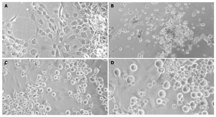
Light-microscopic pictures of cultured cells. Cultured rMSC (A), hepatocyte controls (B), or cocultures of rMSC with hepatocytes (C + enlargement D) after 3 wk. Cultured rMSC grew adherent and adopted a polygonal cell morphology after weeks in culture (A). Hepatocytes formed clumps of rounded cells, and few viable cells were observed after 3 wk in culture (B). In mixed cultures, mainly MSC attached to the substratum, whereas hepatocytes grew over the MSC-layer forming clusters (C). Binucleated cells were found within the attached layer of the cultured MSC beginning with 1 wk in culture. Shown is one example at wk 2 out of three experiments. Original magnification ×200.