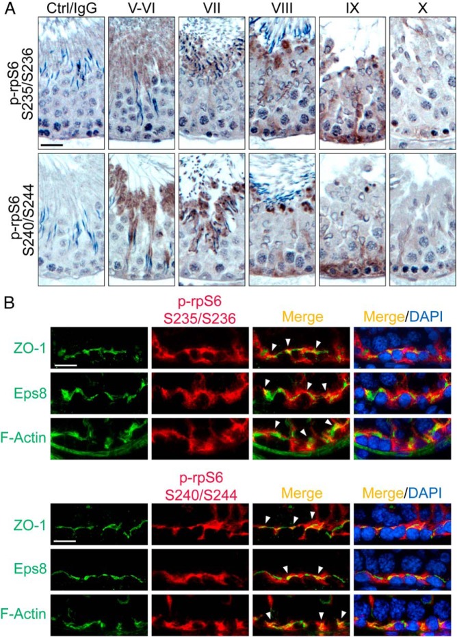
Figure 1.
Immunohistochemical localization of p-rpS6-S235/S236 and p-rpS6-S240/S244 in adult rat testes. A, Using paraffin sections, localization of p-rpS6-Ser235/Ser236 (red fluorescence) vs p-rpS6-Ser240/Ser244 (red fluorescence) in the seminiferous epithelium was examined, in which p-rpS6 appeared as brownish precipitates near the basement membrane, consistent with their localization at the BTB. Phosphorylation of rpS6 at these 4 sites appeared at stage VIII and became more robust at stage IX but considerably diminished by stage X. Scale bar, 50 μm. Control (Ctrl) shown in first column is the cross-section of a testis in which the primary antibody was substituted by normal rabbit IgG, illustrating the staining specificity shown in other micrographs herein. B, Frozen sections were used to confirm their colocalization with BTB proteins ZO-1 (green fluorescence) and Eps8 (green fluorescence), and also F-actin (green fluorescence), by dual-labeled immunofluorescence analysis in which colocalized proteins (yellowish-orange to orange fluorescence) were annotated by white arrowheads. Nuclei were stained with DAPI (blue). Scale bar, 25 μm. S, Ser.

