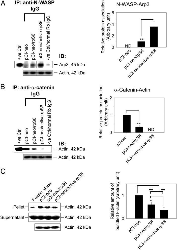
Figure 4.
Overexpression of quadruple rpS6 phosphomimetic mutant in Sertoli cells shifted actin cytoskeleton from bundled to branched configuration mediated by Arp3. Sertoli cells cultured for 2 days with an established TJ barrier were transfected with corresponding vector for 18 hours. Two days after transfection, cells were harvested to obtain lysates for Co-IP and actin bundling assay. A, Co-IP that accessed protein-protein association of N-WASP and Arp3 was performed using Sertoli cell lysates (left panel). Cell lysates (∼400-μg protein) incubated with normal rabbit IgG instead of precipitating antibody served as a negative control. Lysate (5-μg protein) from cells transfected with empty vector without Co-IP served as a positive control with β-actin as the protein loading control. Data are representative results of n = 4 experiments. A histogram (right panel) summarizing Co-IP results shown on the left panel. Each data point was first normalized against the corresponding actin level and then against the protein-protein interaction level in pCI-neo (control) or pCI-neo/rpS6, which was arbitrarily set at 1. Each bar is a mean ± SD of 4 independent experiments. **, P < .01. ND, not detectable because the level of a target protein was too low or it could not be consistently detected in multiple experiments. B, Co-IP that assessed changes in the association between α-catenin and actin using Sertoli cell lysates (left panel) under the experimental conditions as described in A. A histogram (right panel) summarizes Co-IP results from 4 experiments. ND, not detectable. **, P < .01. C, Lysate obtained from Sertoli cells transfected with different corresponding vector was assessed for actin bundling activity (left panel). Linear and unbundled actin filaments were found in supernatant whereas bundled F-actin were sedimented in the pellet after centrifugation (see Materials and Methods). Free F-actin without incubation with cell lysates (F-actin alone) served as a negative control, illustrating that Sertoli cell lysate was capable of bundling actin microfilaments. Data are representative findings of 4 experiments. A histogram (right panel) summarizes results shown in the left panel. Each data point was normalized against the corresponding actin level and then against the protein level in pCI-neo, which was arbitrarily set at 1. Each bar is a mean ± SD of 4 independent assays. *, P < .05; **, P < .01.

