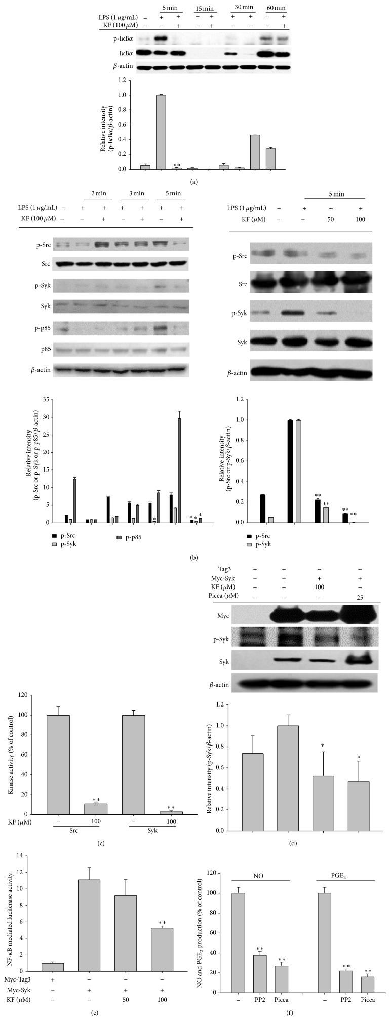
Figure 3.
The effects of KF on NF-κB activation signaling. (a and b) RAW264.7 cells (5 × 10⁶ cells/mL) were incubated with LPS (1 μg/mL) in the presence or absence of KF for the indicated times. After preparing the whole lysates, the levels of total or phosphorylated IκBα, Src, Syk, and p85 were identified using immunoblot analyses. (c) The inhibitory effects of KF on Src and Syk activity were determined using a conventional kinase assay with purified Src and Syk. (d) HEK293 cells transfected with Myc-Syk cDNA (1 μg/mL) for 24 h were treated with KF or Picea for 12 h. After preparing the whole lysates, the levels of total or phosphorylated Myc, Syk, and β-actin were identified using immunoblot analyses. (e) HEK293 cells cotransfected with the NF-κB-Luc (1 μg/mL each) and β-gal (as a transfection control) plasmid constructs were treated with KF in the presence or absence of Myc-Syk for 12 h. Luciferase activity was determined using luminometry. (f) The inhibitory effects of PP2 or Picea on the production of NO or PGE2 were examined using the Griess assay and EIA. Relative intensity was calculated using total levels by the DNR Bio-Imaging System. All of the data are expressed as the mean ± SD of experiments that were performed with six or three (a, b, c, and d) samples. ∗ P < 0.05 and ∗∗ P < 0.01 compared to the control group.

