
Figure 1.
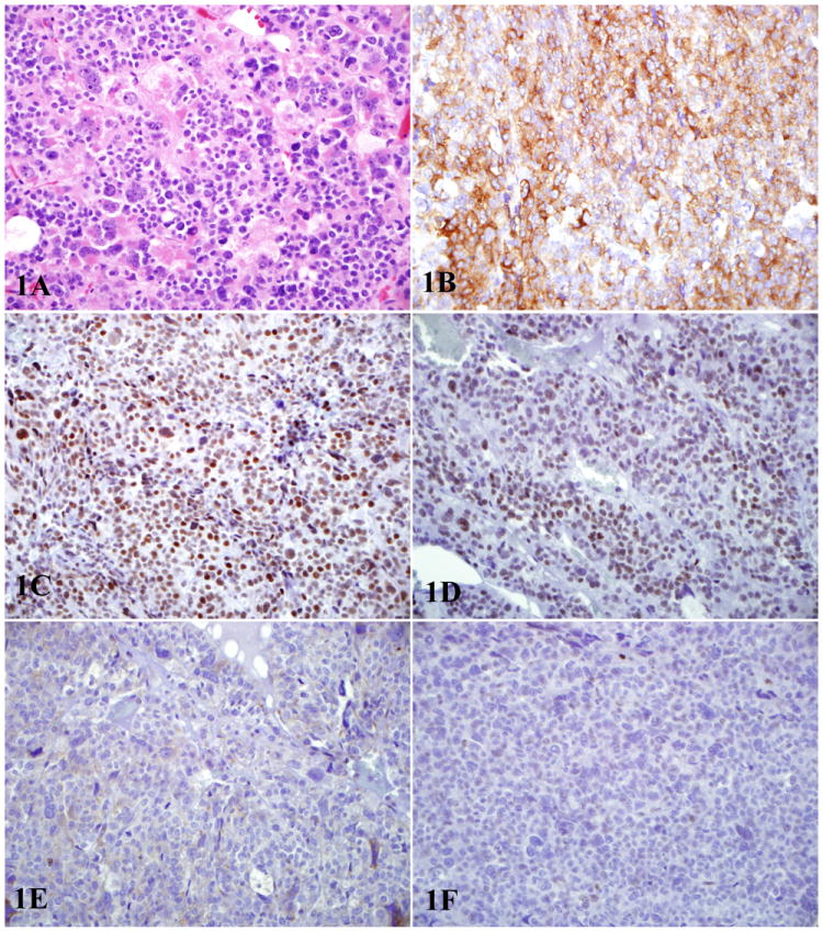
1A: Hematoxylin and eosin stained section of pheochromocytoma with marked cytologic atypia 1B: Tumor shows diffuse cytoplasmic staining for synaptophysin 1C: Tumor demonstrates presence of mismatch repair proteins MLH-1 1D: Tumor demonstrates presence of PMS-2 1E: Tumor shows loss of mismatch repair proteins MSH-2 (1E) 1F: Tumor shows loss of mismatch repair proteins MSH-6 (1F).
* All photos at 400X.