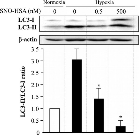
Fig 2.

Western blot analysis of the cytosolic form of antimicrotubule-associated protein 1 light chain 3 (LC3-I) and the LC3–phosphatidylethanolamine conjugate (LC3-II) and the inhibitory activity of nitric oxide donor S-nitrosated human serum albumin (SNO-HSA). Colon cancer C26 cells were incubated under hypoxic conditions with 0.5 and 500 nM SNO-HSA for 24 h. The cell lysates were used to carry out Western blotting for LC3 and β-actin. The density of the bands was quantitatively analyzed using NIH Image J Software and they were standardized by β-actin. Results are given as the means ± SD. *P < 0.05 versus hypoxia group (n = 4).
