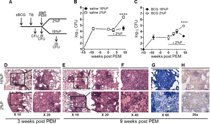
FIG 1.
PEM induces disease relapse in steady-state TB infections in both saline-treated and BCG-vaccinated mice. Mice were vaccinated with BCG or given saline 6 weeks prior to M. tuberculosis infection by the aerosol route. PEM was initiated at week 15 p.i. Lungs were harvested at various time points p.i. and after PEM for assessment of immune responses (IR) and bacterial numbers (CFU). (A) Schematic drawing of the experimental setup (see Materials and Methods for more details). For determination of bacterial loads, lungs were harvested, homogenized, and cultured for 2 to 3 weeks at 37°C before enumeration of bacteria. (B) CFU counts in TB-infected control animals with a normal diet (16%) and PEM (2%). Data are displayed as the mean ± the standard error of the mean (n = 5 or 6 mice per time point). (C) CFU counts of BCG-vaccinated animals were determined as described above. Data are displayed as the mean ± the standard error of the mean (n = 9 to 19 individual mice per time point) and are from two independent experiments. Statistical analysis was done by two-way ANOVA and a Bonferroni posttest. ****, P < 0.0001. (D to H) Photomicrographs of lung tissue from TB-infected, BCG-vaccinated mice on a normal diet (16%P, top) and PEM (2%P, bottom) groups. These were stained with hematoxylin and eosin (D to F), by the Ziehl-Neelsen method (G), and by immunohistochemistry for iNOS (H). The images shown are representative of lesions observed in four mice in each group. Scale bars, 50 μm.

