FIG 2.
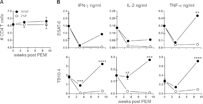
PEM reduces antigen-specific cytokine responses. Groups of mice were vaccinated with BCG, infected with M. tuberculosis, and subsequently fed a 16%P or 2%P diet. Mice were terminated at weeks 0, 3, and 9 after PEM. (A) Lymphocytes from perfused lungs were stained for CD4 and analyzed by flow cytometry. The numbers of CD4 T cells were calculated by multiplying the percentage of CD4 cells (of the total cells in a forward scatter-side scatter plot) by the cell count from the single-cell suspensions for 2%P-fed (open circles) and 16%P-fed (closed circles) mice. Data are expressed as the mean ± the standard error of the mean (n = 4 to 6 mice per group per time point). (B) Lung lymphocytes were harvested from perfused lungs and stimulated with TB10.4 or ESAT-6 in cell culture plates for 72 h at 37°C. The production of IFN-γ, TNF-α, and IL-2 was assessed with an MSD multiplex cytokine kit. Each data point represents a pool of four mice, assessed in technical duplicates or triplicates and represents the mean ± the standard error of the mean. The results shown are representative of two independent experiments. Statistical analysis was done by two-way ANOVA and Bonferroni's multiple-comparison posttest. *, P < 0.05; **, P < 0.01; ***, P < 0.001; ****, P < 0.0001.

