FIG 3.
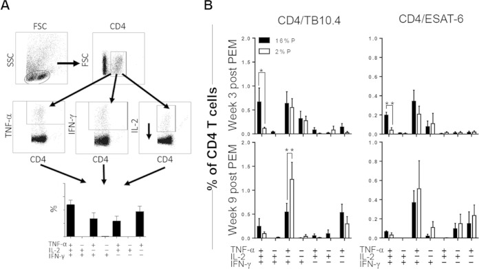
PEM is associated with a reduced proportion of multifunctional CD4 T cells. Groups of mice were vaccinated with BCG, infected with M. tuberculosis, and subsequently fed a 16%P (n = 4 to 7 per time point) or a 2%P (n = 3 to 6 per time point) diet. Mice were sacrificed at weeks 3 (prelapse) and 9 (postlapse) after PEM. Single-cell suspensions obtained from perfused lungs were stimulated in vitro with no antigen, TB10.4, or ESAT-6 for 5 h prior to staining for CD4, TNF-α, IFN-γ, and IL-2 for multicolor ICS flow cytometric analysis. (A) Gating sequence for flow analysis. Lymphocytes were gated from the side scatter (SSC)-forward scatter (FSC) plot, followed by gating of IFN-γ-, TNF-α-, and IL-2-positive cells from the CD4-positive population. With FlowJo for Boolean gating analysis, the antigen-specific CD4 T cells were subdivided into seven distinct subsets based on their cytokine profiles. (B) The ICS data were analyzed as described in Materials and Methods, and the frequency of each CD4 subset is depicted as the mean ± the standard error of the mean for 16%P (black bars) and 2%P (white bars) mice. The results shown are from one experiment and are representative of two individual experiments. All of the background from medium-stimulated cells (<0.5%) has been subtracted. Statistical analysis was done by t test. *, P < 0.05; **, P < 0.01.

