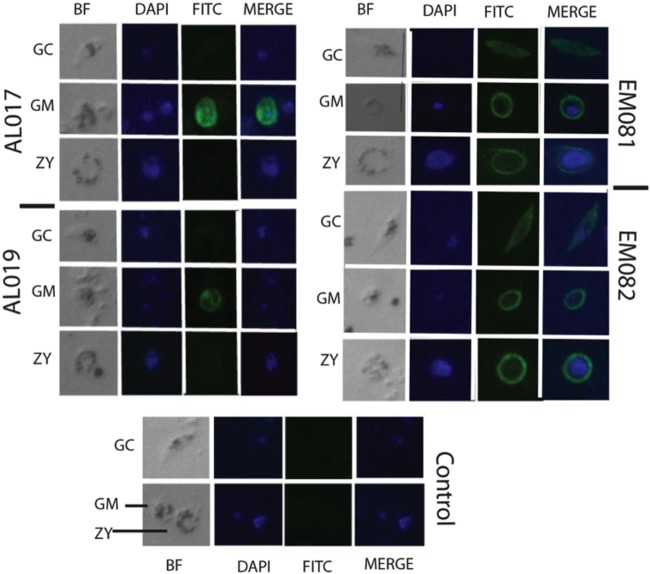
FIG 7.
Immunofluorescence microscopy images show the binding of anti-alga Pfs25 antiserum to native parasite protein expressed in the in vitro-cultured gametocyte (GC), female gamete (GM), and zygote (ZY) stages of P. falciparum. BF, Nomarski (differential interference contrast) bright-field images; DAPI, nucleus stained blue; FITC (fluorescein isothiocyanate), Pfs25 protein stained green; Merge, merged images to show both the nucleus and the Pfs25 protein localization in P. falciparum parasite stages. Serum samples from rPfs25 antigens in the four vaccine groups were used as primary antibodies for probing the specimens.

