
Figure 1.
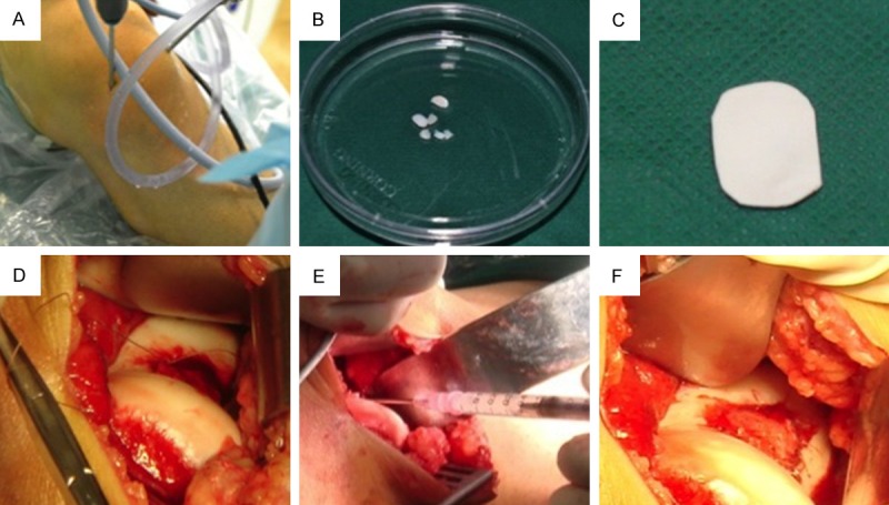
Autologous chondrocyte implantation. A. Cartilage biopsy was harvested from a non-weight bearing area of the knee joint under an arthoscope. B. Around 100 mg cartilage was harvested and enzymatically digested. Chondrocytes were propagated in a GMP lab. C. During the second operation, a collagen membrane was trimmed according to the defect size. D. The periosteum was sutured. E. Chondrocytes were injected into the space between the membrane and the defect. F. After chondrocyte injection, the membrane was sealed with fibrin glue to prevent leaking.