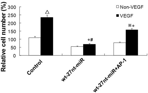
Figure 6.

Analysis of endothelial cell proliferation after VEGF stimulation and AP-1 transfection. Endothelial cells were stimulated with or without VEGF for 24 h. Then cell proliferation of cells transfected with wt-27nt-miR and wt-27nt-miR+AP-1 was analyzed by MTT assay. In control group, compared to control cells without VEGF treatment, ΔP < 0.01. In wt-27nt-miR+AP-1 group, compared to cells without VEGF treatment, ※P < 0.01. Compared to control cells with VEGF treatment, *P < 0.01. Compared to cells in wt-27nt-miR+AP-1 group with VEGF treatment, #P < 0.01.
