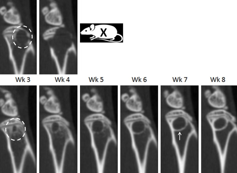
Figure 2.

Micro CT analysis of progressive bone lesion in mouse femur following intracardiac injection. Identical sagittal sections from the same mouse containing a progressive bone lesion from week 3 to week 4 (top panel, Wk3-Wk4) and week 3 to week 8 (bottom panel, Wk3-Wk8). Aggressive lytic lesions appeared within the majority of animals by week 3 (top panel, Wk3, white circle) and breached the cortical bone by week 4 (top panel, Wk4), resulting in removal from the study (top panel, Wk5, X). In a minority of animals, a circular type lytic lesion increased from week 3 to week 6 (bottom panel, Wk3-Wk6). New bone growth (bottom panel, Wk7, white arrow) was first observed prominently at week 6, followed by cessation of lesion progression during week 6 to week 8.
