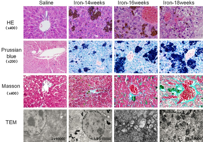
Figure 2.

Histology of liver tissue in gerbils treated with saline or iron dextran for 14, 16 or 18 weeks. Tissues were stained as described in Figure 1. HE staining showed normal hepatocyte structure and well-ordered hepatic cords in the saline group; iron overload tissue showed hepatocyte swelling, fatty changes, soluble cytoplasm, karyopyknosis and disordered hepatic cords. Prussian-blue staining showed no obvious iron deposition in the saline group, compared to abundant single deposits or deposits in clusters, which were observed primarily in Kupffer cells, hepatocytes and macrophages. Masson staining showed minimal fibrosis in blood vessels in saline animals, compared to obvious, extensive liver fibrosis in iron overload animals. TEM revealed normal organelle structure in saline animals, compared to abundant iron deposits (possibly in lysosomes) around the nucleus, together with mitochondrial swelling and vacuolization.
